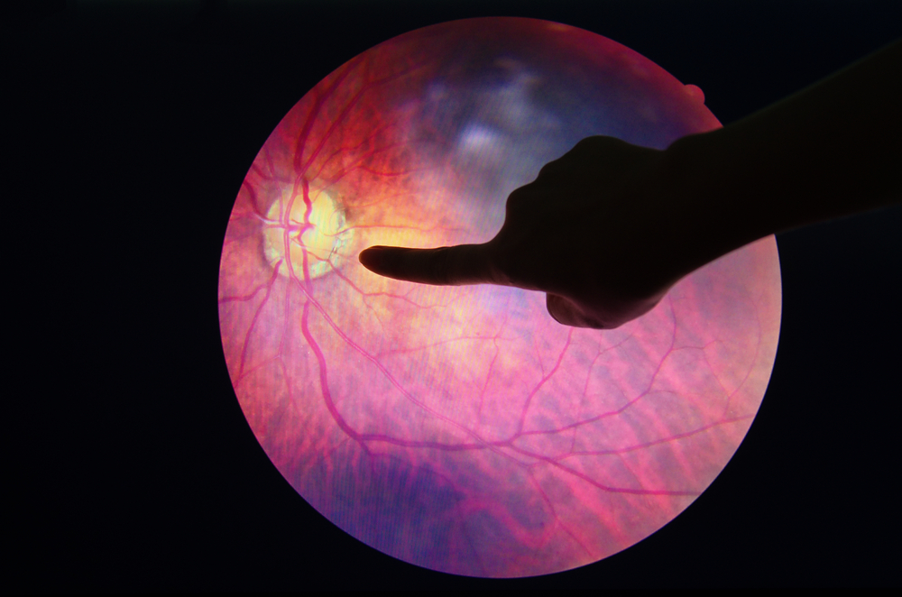
Don’t Let Glaucoma Rob You of Your Eyesight!

The eye is one of life’s treasures. It is our organ for vision.

The retina, located at the back of our eye, plays a key role in giving us our gift of sight. This is where information on shape, colour and pattern is picked up and carried to the brain via the optic nerve.

We must, therefore, be vigilant of eye conditions that may damage our optic nerve, impact our vision and eventually lead to sight loss. One such chronic condition is glaucoma.

WHAT IS GLAUCOMA?

Glaucoma occurs when the optic nerve connecting the eye to the brain is damaged. It often develops because of fluid building up in the in the front part of the eye, resulting in raised intraocular pressure (IOP).

While anyone can develop glaucoma, it is more common in people over 60 years old and can result in vision loss and eventual blindness, if not treated.

With no known cure for glaucoma, regular eye examinations are therefore, very important for early detection and proper treatment to slow or stop its progression.

WHAT ARE SOME SIGNS AND SYMPTOMS?

Symptoms can start so slowly that you may not even notice it.

Thus, it is good to promptly consult your ophthalmologist if you have any tell-tale signs.

These include loss of peripheral vision, having tunnel vision, a blurred or hazy vision, experience of halos or glare especially at night and having some eye pain and discomfort.

HOW TO TREAT GLAUCOMA?

The goal in glaucoma treatment is to control IOP, the main risk factor for the development and progression of glaucoma.

The first and most common treatment option is always noninvasive through medications or eye drops.

In Malaysia, a group of medicines called prostaglandin analogues (PGAs) are a common treatment choice because they are effective in lowering pressure in the eye and are easy to use with daily application.

However, it is reported that about 50% of users of PGA eye drops may experience visible changes that occur around the eye including the eyelids, known as prostaglandin-associated periorbitopathy syndrome (PAPS), after over one month.

Common signs of PAPS are sunken eyes or deepening of the upper eyelid sulcus (also known as DUES), excessive eyelash growth, droopy upper eyelid, darkening of skin around the eyes or eyelids, flattening of the lower eyelid bags and/or fat loss around the eye, and eyelids pressing firmly against the eye.

These can adversely affect the quality of life of glaucoma patients causing them to feel gloomy, avoid looking in the mirror, lack confidence to socialise, and interfere with their work.

Beyond these cosmetic changes, PAPS can also have negative impact on treatment outcomes, including unreliable eye pressure measurements and added difficulty and poor outcomes related to glaucoma surgery in the future.

Some patients are known to discontinue their treatment because of these side effects and this can hamper the overall management of their condition.

Nonetheless, the good news is that with advancements in ophthalmology, there are other class of medicines now available to fulfil the unmet needs among glaucoma patients. These newer options can help them be free from PAPS so they can continue treatment without compromising their quality of life.

BE PROACTIVE FOR BETTER VISION

Detect your condition early for better outcomes and consult your ophthalmologist to learn of a suitable eye drop for you.

Remember, regular and frequent eye examinations are essential to monitor the progression of glaucoma and to adjust treatment as needed.

Prevent the risk of vision loss and continue to enjoy the beautiful wonders of creation.

“Don’t Let Diabetes Ruin Your Eyesight!” Warns Expert

WORDS LIM TECK CHOON

FEATURED EXPERT
DR PEH KHAIK KEE
Consultant Ophthalmologist and Vitreoretinal Surgeon
Sunway Medical Centre
DIABETES IS THE MOST COMMON CAUSE OF BLINDNESS AMONG WORKING-AGE ADULTS

“1 in 5 Malaysians is diabetic, which equals to an estimated 4.6 million Malaysians, and diabetic eye disease affects 1 in 3 diabetics. That means about 500,000 of these people will experience vision-threatening eye disease,” says Dr Peh Khaik Kee.

DIABETIC RETINOPATHY IS A DIABETES-LINKED EYE DISEASE THAT CAN LEAD TO VISION LOSS

According to Dr Peh, diabetes affects the eye in several ways:

  • It causes changes to the lens, leading to blurry vision.
  • Increased pressure in the eye can lead to glaucoma.
  • Damages the nerves that control the eye muscles, giving rise double vision.
  • Damages the blood vessels in the retina, leading to diabetic retinopathy.
Overview of diabetic retinopathy. Click on the image for a larger, clearer version.

Dr Peh shares: “Diabetic retinopathy has become of epidemic proportions. A large part of this issue is due to poor awareness. Many people with diabetes do not undergo regular eye exams to check for signs of the condition.”

REGULAR EYE EXAMS ARE IMPORTANT BECAUSE EARLY STAGE DIABETIC RETINOPATHY DOES NOT SHOW ANY SYMPTOMS

Vision loss occurs at late-stage diabetic retinopathy—this is when most people affected by this disease seeks medical help.

Dark spots floating around in the person’s vision (floaters) are a possible symptom of diabetic retinopathy.

Unfortunately, the vision loss is permanent, and at that stage, there is little to be done to improve the person’s eyesight.

On the other hand, should diabetic retinopathy be detected at its early stages, treatment can be prescribed to help slow down or stop the vision loss over time.

“That is where the importance of screening comes in,” Dr Peh said.

EYE SCREENING FOR DIABETIC RETINOPATHY IS MORE COMFORTABLE THESE DAYS

In the past, such screening can involve staring at a bright light for up to 5 minutes for each eye, which can be very uncomfortable for some people.

Dr Peh shares that the eye screening process has improved since. “These days we have ultra-widefield fundus cameras that can capture a single, 150-degree field view of the retina; 3 times more field view than a standard camera.”

He adds that the camera captures images in under 1 minute, with up to 98% accuracy in detecting and grading retina disease when combined with targeted slit-lamp examination.

Thus, the screening process is faster and hence the person undergoing screening will have a more comfortable experience, while at the same time offering a high accuracy rate in detecting diabetic retinopathy.

EYE SCREENING MAKES A DIFFERENCE

“Diabetic eye screening was formally initiated in the UK in 2009, and by 2014, diabetes is no longer the commonest cause of blindness in working adults in the UK. That is the difference that screening makes,” says Dr Peh.

WHO SHOULD GO FOR EYE SCREENING?

Dr Peh encourages those with a family history of diabetes to have their blood sugar screened, and those diagnosed with diabetes should see an ophthalmologist to have their retina examined.

How Intraocular Lens Can Help People with Cataract and Presbyopia

WORDS LIM TECK CHOON

FEATURED EXPERT
DR CHEONG FOOK MENG
Consultant Ophthalmologist
Gleneagles Hospital Kuala Lumpur
CONCERNING CATARACT

Dr Cheong Fook Meng shares that it is normal to develop cataracts as you age.

However, this condition can also be caused by eye injuries as well as certain diseases or medications.

As one’s cataract progresses, the lens in their eyes gradually become hard and cloudy, allowing less light to pass through. This leads to diminished vision and even complete blindness in severe cases.

ABOUT PRESBYOPIA 

“Presbyopia is another condition that occurs naturally as a person ages; your eyes gradually lose the ability to focus on nearby objects,” says Dr Cheong.

With presbyopia, the lens inside the eye progressively loses its flexibility, making it harder for the affected eye to focus the light reflected from objects.

A common sign that someone may have developed this condition is having to hold reading materials at arm’s length to make out what they are reading.

BOTH CAN BE TREATED WITH SURGERY & IMPLANTATION OF INTRAOCULAR LENS

“This surgical treatment allows the removal of the eye’s cloudy natural lens and replacing it with an intraocular lens,” explains Dr Cheong.

The intraocular lens is clear, made to fit one’s eye shape and personalised according to the patient’s condition and needs.

Intraocular lens (labelled as lens in the image). Click to view a larger version of this image.
TYPES OF INTRAOCULAR LENS AVAILABLE IN MALAYSIA
  • Monofocal lenses correct a single range of vision, usually to see distant objects. Additional issues will need to be fixed through other means.
  • Toric lenses are suitable for patients who also have astigmatism but, similar to monofocal lenses, they restore vision for only one area of focus.
  • Multifocal lenses are designed to provide clear vision for distant and near vision.
GOING FURTHER WITH EXTENDED DEPTH-OF-FOCUS INTRAOCULAR LENS  

Extended depth-of-focus intraocular lens delivers an enhanced range of vision with a reduced frequency of glares and halos, regardless of the lighting conditions.

During the day or when driving at night, these lenses deliver great vision and clarity.

For presbyopia

Extended depth-of-focus intraocular lens can be used to correct presbyopia, by creating a single elongated focal point to enhance one’s range of distance for which their eye can see an object clearly.

For cataract

While all lenses can fix cataracts, extended depth-of-focus intraocular lens may improve one’s quality of life.

They provide distant, intermediate (at arm’s length, such as for reading a newspaper or working on a laptop) and functional near vision (up close, such as for reading books and mobile phone screen), with minimum visual disturbances.

They may even decrease a patient’s need to wear glasses after their cataracts have been removed.