WORDS LIM TECK CHOON
 FEATURED EXPERT FEATURED EXPERTDR JOAN GAN CHEAU YAN Consultant General Surgeon (Special Interest in Breast Oncology Surgery) Regency Medical Care Centre |
UNDERSTANDING DUCTAL CARCINOMA IN SITU
- The word ‘ductal’ is a reference to the milk ducts in the breast. Milk ducts are the small tubes that carry milk from the milk-producing glands to the nipple.
- ‘Carcinoma’ is a type of cancer that starts in the cells lining different parts of the body, such as our skin or internal organs.
- The phrase ‘in situ’ is a Latin term that has been incorporated into everyday use. It refers to being ‘in place’ or ‘in its original position.’
Hence, ductal carcinoma in situ, explains Dr Joan Gan Cheau Yan, refers to cancer cells that are found in the lining of the breast milk duct, and remain confined within that location.
DCIS is classified as stage 0 or the earliest form of breast cancer.
THE CANCER CELLS HAVE NOT YET SPREAD OUTSIDE THE MILK DUCTS TO OTHER PARTS OF THE BREAST
Because of its confinement to the milk ducts, Dr Joan shares that surgery (with or without radiotherapy and/or endocrine therapy) can be very effective.
“If treatment goes well, then chemotherapy is typically not needed,” she says.

Click on the above image for a larger, clearer version.
IF LEFT UNTREATED, CAN THE CANCER CELLS SPREAD TO OTHER PARTS OF THE BODY?
When cancer cells spread outside of their original location—the milk ducts in the case of DCIS—the cancer is said to have become invasive.
“Not all DCIS progresses to invasive cancer,” Dr Joan points out.
- “However, about 20-30% of DCIS may become invasive without definitive treatment,” she adds.
- The risk becomes higher if DICS is high grade; may go up to 50% in some studies.
Grades of DCIS
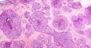
Grading is done when a sample of the cancer cells are examined under the microscope in a histopathology lab.
| Grade 1 (low grade) | The cancer cells still have some resemblance to normal, healthy cells, and tend to grow slowly. |
| Grade 2 (intermediate grade) | The cancer cells have fewer resemblance to healthy cells, and they grow faster than grade 1 cancer cells. |
| Grade 3 (high grade) | The cancer cells look very different from healthy cells and are fast growing. |
A laboratory slide showing the presence of DCIS cells. Click on the image for a larger, clearer version.
HOW CAN ONE TELL WHETHER THEY MAY HAVE DCIS?
- “DCIS usually shows no symptoms (asymptomatic),” Dr Joan tells us.
- However, it may present as painless breast lump in some instances. Hence, regular breast self-examination is useful to detect DCIS and other types of breast cancer.
MAMMOGRAM FOR EARLY DETECTION OF DCIS & OTHER TYPES OF BREAST CANCER
“The majority of DCIS cases are detected during mammogram,” Dr Joan also points out, emphasizing the importance of going for mammogram screening. Suspicious microcalcification is seen during mammogram if DCIS is present.
Dr Joan shares that there is no established method to reduce the risk of developing DCIS. The best recourse we have at the moment is to detect DCIS early via regular mammogram screening, as early detection can improve the odds of a better treatment outcome.
How Does Mammogram Compare to Other Screening Methods?
- Mammogram has a sensitivity of 80% to 95% in detection of breast cancer, the sensitivity is greatly affected by the density of breast tissue.
- Additional ultrasound may sometimes be useful to improve the sensitivity of detection.
- Magnetic resonance imaging (MRI) has the highest sensitivity of detection (up to 95%) but its cost is higher, and its availability can be more limited than mammogram and ultrasound.
RECOMMENDATIONS FOR MAMMOGRAM
|
HOW IS DCIS TREATED?
“Surgery is the mainstay of treatment for current standard of care,” says Dr Joan.
When it comes to breast surgery, the options often include:
- Mastectomy, which removes the breast.
- Breast-conserving surgery, which has become more popular as it allows the patient to keep as much breast tissue as possible.
Breast-Conserving Surgery
“Breast conserving surgery is the preferred method if patient has single focus lesion and the cosmetic outcome is acceptable after the surgery,” says Dr Joan.
Such surgery typically involves the removal of breast lump (lumpectomy).
Dr Joan adds that radiotherapy is usually performed after the surgery to:
- Eliminate any cancer cells that may still be present.
- Reduce the risk of local recurrence in high grade DCIS.
Endocrine therapy may also be prescribed in high-risk cases.
Mastectomy
Mastectomy is the surgical removal of the breast.
“When complete removal (total mastectomy) of the affected breast is needed, axillary lymph node sampling is recommended,” Dr Joan says.
| In lymph node sampling, lymph nodes in fat tissues would be dissected and examined to determine whether cancer cells are present. If cancer cells are present, this suggests that the cancer cells have spread outside their original location. |


