Health Not Sure What Immunotherapy Is? Get Your Answers From an Oncologist! byHealthToday Malaysia15/12/2025
For Her How Combining Anti-Hormone Treatments & CDK4/6 Inhibitors Can Benefit Women with Certain Breast Cancers byHealthToday Malaysia14/12/2025
For Him Men, Outsmart These Two Silent Killer Cancers Before They Strike byHealthToday Malaysia25/11/2025
Health How to Get Free Colorectal Cancer Screening in Malaysia… Before It’s Too Late! byHealthToday Malaysia24/11/2025
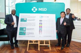
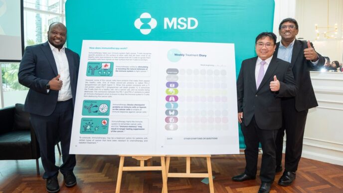
Press Statements Malaysia’s First Lung Cancer Immunotherapy Diary Empowers Patients Every Step of the Way byHealthToday Malaysia18/11/2025
Health How Radiotherapy May Allow Men with Prostate Cancer to Skip Prostate Removal Surgery byHealthToday Malaysia05/11/2025
Health Pink October 2025: The Fight Against Breast Cancer and a New Chapter at Damansara Specialist Hospital byHealthToday Malaysia03/11/2025
For Him How Much Do You Know about Male Breast Cancer? Let’s Find Out! byHealthToday Malaysia24/10/2025
Health Living with a Rare Leukemia: Stories of Struggle, Survival, and Hope byHealthToday Malaysia22/10/2025
Aesthetics Can Cancer Survivors Go for Aesthetic Treatments? Expert Weighs In byHealthToday Malaysia22/10/2025